
Top 10 Chicken Bone X Ray Types for Global Buyers

For global poultry processors, choosing the right chicken bone x-ray system is a practical quality decision, not a simple equipment purchase. Different machines detect different risks. Some identify dense bone fragments inside cooked meat. Others inspect frozen products, whole birds, cartons, or vacuum-packed portions. Image clarity, conveyor speed, product temperature, and software performance all matter. So does the supplier’s technical support.

Dr. Martin Wiedmann, a food-safety professor at Cornell University, has emphasized, “Food safety is a shared responsibility.” That principle fits every chicken bone x-ray application. A processor may install a powerful system, yet still need trained operators, regular calibration, and carefully designed inspection procedures. A bright bone fragment on a monitor can look obvious, but small fragments may hide near joints, packaging folds, or overlapping portions. No machine is flawless. That reality deserves attention.

This guide examines ten chicken bone x-ray types used by buyers across global markets. It considers detection capability, product compatibility, inspection speed, hygiene design, maintenance needs, and total operating value. Some systems suit high-volume poultry lines. Others work better for smaller batches or specialized products. The cheapest option may create higher costs later through downtime, false rejects, or weak service support. Buyers should compare real samples, not only brochures. They should also ask suppliers to explain limitations clearly. A thoughtful selection process can protect consumers, reduce waste, and support more reliable production.
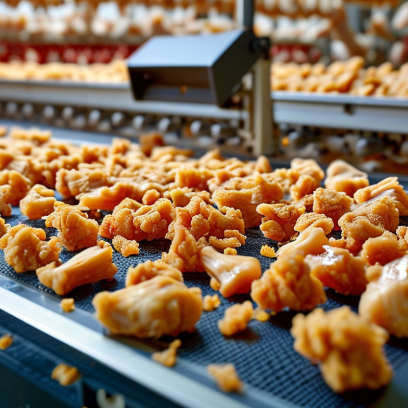
Top 10 Chicken Bone X Ray Types for Global Buyers

Chicken Bone X-Ray Imaging: Purpose, Scope, and Buyer Requirements

Chicken Bone X-Ray Imaging: Purpose, Scope, and Buyer Requirements

Chicken bone X-ray imaging helps processors detect fragments that visual inspection can miss. It supports food safety, quality control, and process improvement. The technology is not a medical test. It evaluates products moving through an industrial inspection line.

The main system types include cabinet, conveyor, portable, line-scan, flat-panel, film, computed radiography, dual-energy, microfocus, and three-dimensional imaging.

Each type serves a different production need. Conveyor systems suit continuous processing. Microfocus systems may reveal smaller fragments. Three-dimensional systems provide deeper inspection, but they usually require higher investment and slower handling.

Conveyor systems suit continuous processing. Microfocus systems may reveal smaller fragments. Three-dimensional systems provide deeper inspection.

Buyers should match the system with product density, package size, line speed, and expected bone dimensions.

Detection performance can change between whole chicken, minced meat, and frozen portions. Ask suppliers to test real samples, including wet packs and overlapping pieces. Request measurable results, not broad claims.

Check image resolution, rejection accuracy, software records, cleaning access, radiation shielding, and operator training. Data export can also support audits and traceability.

Check image resolution, rejection accuracy, software records, cleaning access
Radiation shielding, and operator training
Data export can also support audits and traceability.

Do not judge equipment by sensitivity alone.

A highly sensitive system may create excessive false rejects. That wastes product and time. Trial results can also vary between laboratories and production floors. This is an area where buyers should remain cautious.

Calibration routines, maintenance response, and local compliance documents deserve equal attention. Fancy imaging cannot repair poor product presentation.


Core X-Ray Types for Examining Chicken Bone Structure and Defects

Top 10 Chicken Bone X-Ray Types for Global Buyers

Core X-Ray Types for Examining Chicken Bone Structure and Defects

Global poultry output now exceeds 140 million tonnes annually, according to the FAO Food Outlook 2024. This scale increases demand for reliable bone inspection. The ten core X-ray types include plain film radiography, digital radiography, computed radiography, microfocus radiography, line-scan X-ray, fluoroscopy, tomosynthesis, dual-energy X-ray, cone-beam CT, and micro-CT.

Plain and digital radiography provide fast views of fractures, abnormal density, and incomplete bone removal. Computed radiography remains useful where existing equipment needs upgrading. Microfocus systems reveal fine cracks with smaller focal spots. Line-scan X-ray supports continuous processing. It needs careful calibration.

Fluoroscopy shows movement during positioning, while tomosynthesis creates layered images from limited angles. Dual-energy systems can improve material separation, although poultry tissue creates difficult overlaps. Cone-beam CT gives three-dimensional views of joints and embedded fragments. Micro-CT offers exceptional detail for laboratory validation, not high-speed production. The IAEA emphasizes exposure optimization, shielding, and routine image-quality checks in its radiation safety guidance. OECD-FAO Agricultural Outlook 2024–2033 also identifies poultry as a continuing growth sector, increasing pressure for stable inspection capacity. No method is perfect. In practice, buyers should compare resolution, conveyor speed, dose control, reject accuracy, and maintenance records rather than trust impressive sample images.

Top 10 Chicken Bone X-Ray Types for Global Buyers - Core X-Ray Types for Examining Chicken Bone Structure and Defects
Rank X-Ray Type Category Primary Examination Purpose Bone Structures Best Evaluated Defects or Findings Commonly Detected Typical Chicken Specimens Main Purchasing Considerations
1 Anteroposterior (AP) or Ventrodorsal (VD) Projection Standard projection Provides the principal frontal image for routine assessment of bone alignment, length, and overall mineralization. Long-bone shafts, joint spaces, cortical margins, and general skeletal symmetry. Fractures, angular deformity, bone shortening, abnormal mineral density, and gross joint displacement. Femur, tibia, tarsometatarsus, wing bones, and whole-bird skeletal surveys. Look for consistent exposure control, a suitable detector area, and positioning accessories for small poultry specimens.
2 Mediolateral or Lateromedial Projection Standard projection Supplies a side view that complements the frontal image and reduces the risk of missing displacement in the depth plane. Bone thickness, cranial and caudal cortices, joint congruity, and fracture angulation. Overlapping fractures, luxation, callus formation, cortical irregularity, and sagittal displacement. Femur, tibia, humerus, radius, ulna, and lower-leg joints. A narrow field of view and accurate collimation are useful for improving contrast and limiting scatter.
3 Oblique Projection Specialized projection Rotates the specimen to separate superimposed structures that may be hidden on AP/VD and lateral views. Articular surfaces, condyles, thin cortical boundaries, and complex portions of the wing and leg. Small fissures, subtle cortical defects, incomplete fractures, mineralized lesions, and joint-surface abnormalities. Elbow, knee, ankle, shoulder, hip, and small bones of the distal limbs. Choose systems that support repeatable positioning angles and stable specimen fixation.
4 Craniocaudal and Caudocranial Projection Joint projection Shows selected joints and limb segments from the cranial or caudal direction for improved assessment of alignment. Joint spaces, trochlear regions, condyles, proximal limb ends, and paired cortical margins. Joint incongruity, epiphyseal irregularity, growth-related deformity, and transverse fracture displacement. Stifle, tarsal joint, shoulder, elbow, and proximal or distal long-bone regions. Useful equipment should allow precise limb positioning and clear imaging of small joint spaces.
5 Horizontal-Beam Lateral Projection Trauma projection Captures a lateral image while the specimen remains supported, helping evaluate fluid levels and displaced fragments. Fracture sites, joint spaces, and soft-tissue margins adjacent to injured bones. Acute fracture displacement, joint effusion, soft-tissue swelling, and gas or fluid interfaces. Traumatized limbs, immobilized specimens, and cases where conventional positioning is impractical. Prioritize adjustable tube-detector geometry, safe specimen support, and reliable low-exposure imaging.
6 Stress or Loaded Projection Functional projection Images a joint or limb under controlled positioning or load to reveal instability that may not appear in a neutral view. Joint alignment, ligament-supported spaces, angular relationships, and weight-bearing segments. Dynamic instability, abnormal joint opening, malalignment, and subtle displacement. Stifle, hock, hip, wing joints, and legs used in biomechanics or veterinary research. Use only with an appropriate positioning fixture and a protocol that applies repeatable, controlled force.
7 Whole-Skeleton Survey Radiography Screening examination Provides a broad overview of the axial and appendicular skeleton for screening and comparative studies. Spine, pelvis, ribs, wings, legs, bone length, and overall skeletal symmetry. Multiple fractures, generalized mineralization changes, severe deformity, developmental abnormalities, and asymmetry. Intact chicken carcasses, research specimens, hatchling studies, and comparative poultry samples. A large detector, uniform exposure, image stitching capability, and a stable flat positioning surface are advantageous.
8 Digital Radiography (DR) Digital detector method Creates an electronic projection image for rapid review, storage, measurement, and image processing. Most chicken bones visible on conventional projection images, including cortical margins and joint regions. Fractures, deformities, radiolucent areas, increased opacity, and changes in bone alignment. Routine laboratory examinations, veterinary diagnostics, quality-control sampling, and research workflows. Evaluate detector size, pixel pitch, exposure latitude, image-processing controls, software compatibility, and service support.
9 Micro-Computed Tomography (Micro-CT) 3D high-detail imaging Generates cross-sectional and three-dimensional images for detailed analysis of internal bone architecture. Trabecular pattern, cortical thickness, medullary cavity, mineralized callus, and complex joint anatomy. Microfractures, porosity, trabecular loss, cortical defects, healing architecture, and small structural lesions. Excised chicken bones, small bone segments, embryos, and fixed research specimens. Consider voxel size, specimen chamber dimensions, reconstruction software, radiation shielding, and ex vivo workflow requirements.
10 Fluoroscopic X-Ray Imaging Real-time imaging Displays continuous or pulsed X-ray images to observe movement, positioning, or contrast-flow behavior in real time. Moving joints, skeletal articulation, alignment during manipulation, and selected dynamic anatomical relationships. Functional instability, abnormal motion, movement-related impingement, and changes that are difficult to interpret from static images. Live poultry research, biomechanical studies, procedural guidance, and controlled motion experiments. Review pulse rate, image intensifier or flat-panel performance, dose management, field size, and operator protection features.
Note: The appropriate X-ray method depends on the specimen size, anatomical region, examination objective, required spatial detail, and whether static or dynamic information is needed.

Advanced Imaging Methods for Fractures, Density, and Foreign Materials

Top 10 Chicken Bone X-Ray Types for Global Buyers

Advanced Imaging Methods for Fractures, Density, and Foreign Materials

Chicken bone inspection now uses more than a single flat image. Standard projection radiography provides a fast view of cracks, bends, and separation points. Digital radiography improves contrast and supports quick image sharing. Dual-energy imaging can help distinguish bone from selected foreign materials. It is useful when metal, glass, or dense fragments may be present. Results still depend on exposure settings and sample positioning.

Micro-CT reveals fine internal structures with detailed cross-sectional slices. Cone-beam CT offers three-dimensional views for complex fractures and overlapping bones. Computed tomography measures density patterns across the sample. These methods can show hollow areas, hidden cracks, and unusual mineral loss. Fluoroscopy records movement during controlled handling. This may expose unstable fragments, although it is less suitable for every inspection line. A calibrated density reference remains important. Without it, comparisons can become subjective.

Ultrasound-assisted imaging may support surface checks, but bone blocks sound waves. It should not replace X-ray methods. High-resolution radiography works well for thin specimens and routine screening. Industrial X-ray laminography can help inspect layered or irregular samples. Image analysis software may flag fracture lines and foreign objects, yet human review remains necessary. Artifacts can resemble cracks. Dense tissue can hide small materials. Even experienced operators may disagree. That weakness deserves attention, especially when buyers compare suppliers across different inspection standards. A documented method, trained reviewer, and retained images create stronger evidence than a single pass-fail result.

Comparing the Top 10 Chicken Bone X-Ray Types by Use and Performance

Chicken bone X-ray systems differ sharply by use and performance. Single-energy 2D scanners suit stable, high-volume lines with moderate sensitivity. High-resolution 2D units reveal small fragments in breast fillets. Dual-energy systems separate bone-like density from food more effectively. Dual-view scanners reduce blind spots around joints and overlapping pieces. Line-scan cameras support continuous conveyor inspection. Area-scan systems fit batch checks and irregular product layouts.

AI-assisted 2D systems improve image classification, but training quality remains critical. CT scanners create layered images and help investigate recurring defects, although they cost more and operate slower. Robotic, targeted X-ray cells work well for premium cuts and sampling stations. Portable units support offline verification, not necessarily full production control. That distinction matters.

FAOSTAT’s 2023 dataset recorded global poultry-meat output above 140 million tonnes in 2022. Such scale increases demand for fast, stable inspection. USDA Food Safety and Inspection Service guidance also emphasizes validated control measures, documented monitoring, and corrective action. Buyers should compare detection limits, conveyor speed, reject accuracy, image archives, and sanitation design. A system detecting a 1.0-millimeter fragment may perform differently on skin-on meat, frozen portions, or overlapping bones. Real factory trials are essential. Laboratory results can mislead.

Performance is never just a sensor number. Calibration drift, vibration, moisture, and product spacing affect results. Even advanced software can miss unusual fragments. That weakness deserves attention. A practical evaluation should test at least three product conditions, record false rejects, and repeat trials across shifts.

Global Buyer Factors: Safety, Compliance, Cost, and Equipment Selection

Top 10 Chicken Bone X-Ray Types for Global Buyers

For global buyers, chicken bone X-ray selection starts with the inspection goal, not the catalogue label. Common categories include whole-carcass, breast, thigh, wing, drumstick, neck, back, minced-bone, processed-meat, and foreign-object inspection. Each sample presents a different density pattern.

Small wing bones may need higher image detail, while packed products need stronger penetration. In my purchasing work, I ask suppliers for images from the buyer’s actual product line. Laboratory samples can look perfect. Production samples are less forgiving.

Safety must be documented through shielding design, leakage testing, warning controls, operator training, and maintenance records. Ask whether the proposed system matches electrical and radiation requirements in the destination market.

Compliance is not a single certificate. Import rules, food procedures, installation permits, and inspection practices may differ.

A credible supplier should provide traceable test data, manuals, and clear service boundaries. I would also verify calibration frequency and reject vague detection claims.

Cost comparisons should include installation, staff training, calibration, downtime, and rejected product disposal. A low purchase price may hide expensive false rejects. Conversely, weak sensitivity can create unsafe confidence.

Select detector size, belt speed, power range, and software functions around real throughput. Test bone fragments in thick packages, condensation, and routine cleaning conditions. This is where assumptions break.

I once treated nominal sensitivity as a final answer; it was not. Buyers should record test conditions, acceptance limits, and follow-up responsibilities before signing.

AVADA IT

Fusce ut ipsum tincidunt, porta nisl sollicitudin, vulputate nunc. Cras commodo leo ac nunc convallis ets efficitur.

RECENT TWEETS

CONTACT US

  • 12345 North Main Street,
    New York, NY 555555

  • 1.800.555.6789

  • sale@vibrotools.com

Go to Top